 |
LigASite database of binding sites |
|
PDB ID and HEADER, TITLE and
COMPND records of the PDB file. | | (click anywhere in this window to remove it) |
|
| 2gub |
|
|
| CRYSTAL STRUCTURE OF METAL FREE D-XYLOSE ISOMERASE. |
TITLE |
|
|
|
|
|
|
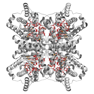
Figure highlighting the binding site residues. Figures were drawn with
Molscript (7) and rendered with
Raster3D (8). PISA coordinates
(3) are used when available
(all entries except NMR). | | (click anywhere in this window to remove it) |
|
 |
|
List of binding site residues detected in this protein. Column 1 gives the position, coloured on a yellow-to-red scale depending on the fraction of corresponding holo-structures where the residue is in contact with a ligand.
Column 2 gives the 3-letter amino acid code, coloured according to physico-chemical type. Chain ID's of residues are not mentioned in this page because all chains in the apo-structure refer to the same protein. | | (click anywhere in this window to remove it) |
|
| 16 | |
TRP |
| 26 | |
PHE |
| 54 | |
HIS |
| 88 | |
MET |
| 90 | |
THR |
| 91 | |
THR |
| 94 | |
PHE |
| 135 | |
VAL |
| 137 | |
TRP |
| 181 | |
GLU |
| 183 | |
LYS |
| 215 | |
ASN |
| 217 | |
GLU |
| 219 | |
GLY |
| 220 | |
HIS |
| | 245 | |
ASP |
| 247 | |
ASN |
| 255 | |
ASP |
| 257 | |
ASP |
| 285 | |
HIS |
| 287 | |
ASP |
| 289 | |
LYS |
|
|
|
| PDB |
The Protein Data Bank |
| CSA |
Catalytic Site Atlas |
| PDBSum |
Overview of the macromolecular structure |
| CATH |
Protein Structure Classification |
| Scop |
Structural Classification of Proteins |
| Pfam |
Protein Families and Domains |
| UniProt |
Universal Protein Resource |
LIGPLOT (only on holo-pages) is hosted at the EBI. The LigPlot Jmol links point directly to the Jmol visualisation interface provided on the PDBSum page. Note that due to different software used, the atomic contacts of LigPlot and LigASite do not necessarily correspond. | | (click anywhere in this window to remove it) |
|
Links to external databases: |
|
Several files are provided for download: | • The XML file defining the residue-ligand contacts; this file contains data on the apo and all holo-structures. |
| • The XML Schema file defining the semantics of the XML file |
| • 3D coordinates of the structure used in constructing LigASite (PISA structure file whenever available, PDB file otherwise. |
| • 3D coordinates of the combined binding residues in the apo structure |
| • 3D coordinates of the binding residues of the holo structure (only on the holo page) |
Coordinate files are in PDB format. | | (click anywhere in this window to remove it) |
|
|
|
|
|
Table describing the holo-structures and ligands used to define
the binding sites.
Column 1 gives the PDB ID of the holo-structure.
Column 2 gives the unique ID of the ligand;
a space-separated list of HET-groups that constitute
the ligand (see Methods).
Each HET-group in the ligand is uniquely identified by
a string in which the first four characters are the three-letter
HET ID from the PDB file followed by the chain ID from
the PISA file, and the last four characters are the residue sequence
number from the PDB file.
Column 3 gives the number of atoms in each ligand.
Column 4 gives the number of protein-ligand inter-atomic
contacts. | | (click anywhere in this window to remove it) |
|
| pdb ID |
Ligand Unique ID |
#atoms |
#contacts |
| 3kbn |
GLOO_401 _NIM_391 _NIN_393 _NIL_392 |
15 |
81 |
Details |
|
GLOT_401 _NIQ_392 _NIS_393 _NIR_391 |
15 |
81 |
|
GLOJ_401 _NIG_392 _NIH_391 _NII_393 |
15 |
80 |
|
GLOE_401 _NIB_392 _NIC_391 _NID_393 |
15 |
80 |
| 3kco |
GLOS_401 _NIT_393 _NIQ_392 _NIR_391 |
26 |
81 |
Details |
|
GLON_401 _NIO_393 _NIM_391 _NIL_392 |
26 |
81 |
|
GLOI_401 _NIG_392 _NIJ_393 _NIH_391 |
26 |
80 |
|
GLOD_401 _NIE_393 _NIB_392 _NIC_391 |
26 |
80 |
| 3kcl |
GLCH_401 _CDG_392 _CDF_391 |
26 |
80 |
Details |
|
GLCP_401 _CDN_391 _CDO_392 |
26 |
80 |
|
GLCL_401 _CDK_392 _CDJ_391 |
26 |
80 |
|
GLCD_401 _CDB_391 _CDC_392 |
26 |
80 |
| 3kbm |
GLCH_401 _CDG_392 |
13 |
73 |
Details |
|
GLCP_401 _CDO_392 |
13 |
70 |
|
GLCD_401 _CDC_392 |
13 |
73 |
|
GLCL_401 _CDK_392 |
13 |
70 |
| 3cwh |
XULG_401 _MGI_392 _OHJ_501 _MGH_391 |
23 |
75 |
Details |
|
XULL_401 _MGN_392 _MGM_391 _OHO_501 |
23 |
79 |
|
XULQ_401 _OHT_501 _MGS_392 _MGR_391 |
23 |
79 |
|
XULB_401 _MGC_391 _MGD_392 _OHE_501 |
23 |
75 |
|
|
|
|
v9.7
April 2012 |
Interdisciplinary Research Institute, Computational Biology, Villeneuve d'Ascq, France
University College London, Biomolecular Structure and Modelling Unit, London, UK
Hospital for Sick Children and University of Toronto, Structural Biology and Biochemistry Program, Toronto, Canada |
| Script execution time: 0.0651 seconds |