 |
LigASite database of binding sites |
|
PDB ID and HEADER, TITLE and
COMPND records of the PDB file. | | (click anywhere in this window to remove it) |
|
| 2au8 |
|
|
| CATALYTIC INTERMEDIATE STRUCTURE OF INORGANIC PYROPHOSPHATASE |
TITLE |
|
|
| INORGANIC PYROPHOSPHATASE |
COMPND |
|
|
|
|
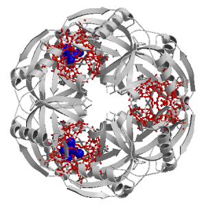
Figure showing the binding site residues. Ligands are displayed as
CPK. Figures were drawn with
Molscript (7) and rendered with
Raster3D (8). PISA coordinates
(3) are used when available
(all entries except NMR). Ligands do not appear on the picture when
PISA fails to apply symmetry operations to ligand coordinates. | | (click anywhere in this window to remove it) |
|
 |
|
List of binding site residues detected in this holo-structure. Column 1 gives the position, coloured on a yellow-to-red scale depending on the number of holo-structures where the residue is in contact with a ligand.
Column 2 gives the identifier of the chain to which the residue belongs.
Column 3 gives the 3-letter amino acid code, coloured according to physico-chemical type. The binding residues in this holo structure are listed for each ligand independently, following the ligand's unique ID. Note that since PISA files are used whenever available, chain identifiers may differ from those in original PDB files. | | (click anywhere in this window to remove it) |
|
| MN R 202 |
|---|
| 20 | K |
GLU |
| 29 | K |
LYS |
| 31 | K |
GLU |
| 55 | K |
TYR |
| 56 | K |
GLY |
| 57 | K |
TYR |
| 67 | K |
ASP |
| 68 | K |
PRO |
| 70 | K |
ASP |
| MN V 202 |
|---|
| 20 | O |
GLU |
| 29 | O |
LYS |
| 31 | O |
GLU |
| 55 | O |
TYR |
| 56 | O |
GLY |
| 57 | O |
TYR |
| 67 | O |
ASP |
| 68 | O |
PRO |
| 70 | O |
ASP |
| PO4 P 180 |
|---|
| 29 | K |
LYS |
| 31 | K |
GLU |
| 43 | K |
ARG |
| 55 | K |
TYR |
| | 65 | K |
ASP |
| 67 | K |
ASP |
| 70 | K |
ASP |
| 102 | K |
ASP |
| 104 | K |
LYS |
| MN T 204 |
|---|
| 29 | K |
LYS |
| 31 | K |
GLU |
| 42 | K |
ASP |
| 43 | K |
ARG |
| 65 | K |
ASP |
| 67 | K |
ASP |
| PO4 T 180 |
|---|
| 29 | O |
LYS |
| 31 | O |
GLU |
| 43 | O |
ARG |
| 55 | O |
TYR |
| 65 | O |
ASP |
| 67 | O |
ASP |
| 70 | O |
ASP |
| 102 | O |
ASP |
| 104 | O |
LYS |
| MN X 204 |
|---|
| 29 | O |
LYS |
| 31 | O |
GLU |
| | 42 | O |
ASP |
| 43 | O |
ARG |
| 65 | O |
ASP |
| 67 | O |
ASP |
| MN Q 203 |
|---|
| 43 | K |
ARG |
| 65 | K |
ASP |
| 97 | K |
ASP |
| 99 | K |
ALA |
| 102 | K |
ASP |
| 141 | K |
TYR |
| 142 | K |
LYS |
| MN U 203 |
|---|
| 43 | O |
ARG |
| 65 | O |
ASP |
| 97 | O |
ASP |
| 99 | O |
ALA |
| 102 | O |
ASP |
| 141 | O |
TYR |
| 142 | O |
LYS |
| CL O 208 |
|---|
| 55 | K |
TYR |
| 95 | K |
MET |
| 97 | K |
ASP |
| 102 | K |
ASP |
| | 104 | K |
LYS |
| 138 | K |
PHE |
| 141 | K |
TYR |
| 142 | K |
LYS |
| CL S 208 |
|---|
| 55 | O |
TYR |
| 95 | O |
MET |
| 97 | O |
ASP |
| 102 | O |
ASP |
| 104 | O |
LYS |
| 138 | O |
PHE |
| 141 | O |
TYR |
| 142 | O |
LYS |
| MN S 201 |
|---|
| 63 | K |
SER |
| 65 | K |
ASP |
| 68 | K |
PRO |
| 70 | K |
ASP |
| 102 | K |
ASP |
| MN W 201 |
|---|
| 63 | O |
SER |
| 65 | O |
ASP |
| 68 | O |
PRO |
| 70 | O |
ASP |
| 102 | O |
ASP |
|
|
|
| PDB |
The Protein Data Bank |
| CSA |
Catalytic Site Atlas |
| PDBSum |
Overview of the macromolecular structure |
| CATH |
Protein Structure Classification |
| Scop |
Structural Classification of Proteins |
| Pfam |
Protein Families and Domains |
| UniProt |
Universal Protein Resource |
LIGPLOT (only on holo-pages) is hosted at the EBI. The LigPlot Jmol links point directly to the Jmol visualisation interface provided on the PDBSum page. Note that due to different software used, the atomic contacts of LigPlot and LigASite do not necessarily correspond. | | (click anywhere in this window to remove it) |
|
Links to external databases: LigPlot (hosted at the EBI):
|
|
Several files are provided for download: | • The XML file defining the residue-ligand contacts; this file contains data on the apo and all holo-structures. |
| • The XML Schema file defining the semantics of the XML file |
| • 3D coordinates of the structure used in constructing LigASite (PISA structure file whenever available, PDB file otherwise. |
| • 3D coordinates of the combined binding residues in the apo structure |
| • 3D coordinates of the binding residues of the holo structure (only on the holo page) |
Coordinate files are in PDB format. | | (click anywhere in this window to remove it) |
|
|
|
|
|
List of related structure, containing both the apo-structure
and other holo-structures.
Column 1 gives the PDB ID and column 2 the unique ID
of the ligands (holo-structures only).
Clicking the blue 'Hide table of related structures' button
removes the entire table. | | (click anywhere in this window to remove it) |
|
|
| pdb ID |
|---|
| 1igp |
|
Details |
|
| pdb ID |
Ligand Unique ID |
|---|
| 2au6 |
POPT_182 _MNS_203 __FX_211 _MNV_201 _MNU_202 |
Details |
|
POPP_182 _MNQ_202 _MNR_201 __FT_211 _MNO_203 |
|
POPH_182 _MNG_203 _MNI_202 __FL_211 _MNJ_201 |
|
POPR_182 _MNQ_203 _MNS_202 _MNT_201 __FV_211 |
|
POPF_182 _MNG_202 _MNH_201 __FJ_211 _MNE_203 |
|
POPD_182 __FH_211 _MNE_202 _MNC_203 _MNF_201 |
| 1i6t |
POPD_411 _CAZ_302 _CAB_303 _CAA_301 |
Details |
|
POPX_411 _CAV_303 _CAU_301 _CAT_302 |
|
POPT_411 _CAP_302 _CAR_303 _CAQ_301 |
|
POPJ_411 _CAH_303 _CAG_301 _CAF_302 |
|
POPH_411 _CAF_303 _CAE_301 _CAD_302 |
|
POPN_411 _CAL_303 _CAK_301 _CAJ_302 |
| 2auu |
POPG_180 _MGE_201 __FH_211 _MGF_204 _MGC_202 _MGD_203 |
Details |
|
POPI_180 __FJ_211 _MGE_202 _MGH_204 _MGG_201 _MGF_203 |
|
POPW_180 _MGV_204 _MGS_202 __FX_211 _MGT_203 _MGU_201 |
|
POPC_180 _MGZ_203 _MGY_202 __FD_211 _MGB_204 _MGA_201 |
|
POPS_180 _MGP_203 __FT_211 _MGQ_201 _MGO_202 _MGR_204 |
|
POPM_180 __FN_211 _MGK_201 _MGI_202 _MGJ_203 _MGL_204 |
| 2au9 |
POPS_180 _MNU_204 _MNQ_203 _MNT_201 _MNR_202 _CLP_208 __FV_211 |
Details |
|
POPH_180 _MNI_201 _CLE_208 _MNG_202 _MNJ_204 __FK_211 _MNF_203 |
|
POPO_180 _MNM_203 _MNQ_204 _MNP_201 _MNN_202 _CLL_208 __FR_211 |
|
POPK_180 _MNI_203 _CLH_208 _MNL_201 _MNJ_202 __FN_211 _MNM_204 |
|
POPD_180 _MNC_202 _MNE_201 _CLA_208 _MNF_204 _MNB_203 __FG_211 |
|
POPZ_180 _CLW_208 _MNB_204 __FC_211 _MNX_203 _MNA_201 _MNY_202 |
|
|
|
Ligands present in this holo structure.
Column 1 shows the ligand HET code
Column 2 shows the name, chemical formula and (non-stereo) SMILES string.
Data in column 2 appears as 'not_found' when it is not present in the file
'pdb2smiles.xml' from www.rcsb.org. | | (click anywhere in this window to remove it) |
|
| PO4 |
NAME: |
PHOSPHATE ION |
|
FORMULA: |
O4 P1 |
|
SMILES: |
[O-]P([O-])([O-])=O |
| MN |
NAME: |
MANGANESE (II) ION |
|
FORMULA: |
MN1 |
|
SMILES: |
[Mn++] |
| CL |
NAME: |
CHLORIDE ION |
|
FORMULA: |
CL1 |
|
SMILES: |
[Cl-] |
|
|
|
|
v9.7
April 2012 |
Interdisciplinary Research Institute, Computational Biology, Villeneuve d'Ascq, France
University College London, Biomolecular Structure and Modelling Unit, London, UK
Hospital for Sick Children and University of Toronto, Structural Biology and Biochemistry Program, Toronto, Canada |
| Script execution time: 1.7636 seconds |