 |
LigASite database of binding sites |
|
PDB ID and HEADER, TITLE and
COMPND records of the PDB file. | | (click anywhere in this window to remove it) |
|
| 3l2d |
|
|
| GLYCOCYAMINE KINASE, BETA-BETA HOMODIMER FROM MARINE WORM NAMALYCASTIS SP. |
TITLE |
|
|
| GLYCOCYAMINE KINASE BETA CHAIN |
COMPND |
|
|
|
|
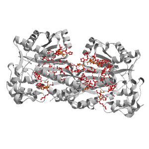
Figure highlighting the binding site residues. Figures were drawn with
Molscript (7) and rendered with
Raster3D (8). PISA coordinates
(3) are used when available
(all entries except NMR). | | (click anywhere in this window to remove it) |
|
 |
|
List of binding site residues detected in this protein. Column 1 gives the position, coloured on a yellow-to-red scale depending on the fraction of corresponding holo-structures where the residue is in contact with a ligand.
Column 2 gives the 3-letter amino acid code, coloured according to physico-chemical type. Chain ID's of residues are not mentioned in this page because all chains in the apo-structure refer to the same protein. | | (click anywhere in this window to remove it) |
|
| 82 | |
LYS |
| 83 | |
LYS |
| 84 | |
THR |
| 85 | |
GLY |
| 139 | |
SER |
| 140 | |
CYS |
| 141 | |
ARG |
| 143 | |
ARG |
| 199 | |
ILE |
| 202 | |
HIS |
| 204 | |
LEU |
| 205 | |
PHE |
| 206 | |
GLU |
| 212 | |
LEU |
| 239 | |
TRP |
| | 242 | |
GLU |
| 243 | |
GLU |
| 247 | |
ARG |
| 251 | |
MET |
| 252 | |
GLN |
| 294 | |
CYS |
| 296 | |
THR |
| 297 | |
ASN |
| 303 | |
ARG |
| 305 | |
SER |
| 306 | |
VAL |
| 307 | |
HIS |
| 331 | |
ARG |
| 333 | |
THR |
| 334 | |
GLY |
| | 335 | |
GLY |
| 336 | |
GLU |
| 337 | |
SER |
| 346 | |
ASP |
| 350 | |
TRP |
| 352 | |
ARG |
|
|
|
| PDB |
The Protein Data Bank |
| CSA |
Catalytic Site Atlas |
| PDBSum |
Overview of the macromolecular structure |
| CATH |
Protein Structure Classification |
| Scop |
Structural Classification of Proteins |
| Pfam |
Protein Families and Domains |
| UniProt |
Universal Protein Resource |
LIGPLOT (only on holo-pages) is hosted at the EBI. The LigPlot Jmol links point directly to the Jmol visualisation interface provided on the PDBSum page. Note that due to different software used, the atomic contacts of LigPlot and LigASite do not necessarily correspond. | | (click anywhere in this window to remove it) |
|
Links to external databases: |
|
Several files are provided for download: | • The XML file defining the residue-ligand contacts; this file contains data on the apo and all holo-structures. |
| • The XML Schema file defining the semantics of the XML file |
| • 3D coordinates of the structure used in constructing LigASite (PISA structure file whenever available, PDB file otherwise. |
| • 3D coordinates of the combined binding residues in the apo structure |
| • 3D coordinates of the binding residues of the holo structure (only on the holo page) |
Coordinate files are in PDB format. | | (click anywhere in this window to remove it) |
|
|
|
|
|
Table describing the holo-structures and ligands used to define
the binding sites.
Column 1 gives the PDB ID of the holo-structure.
Column 2 gives the unique ID of the ligand;
a space-separated list of HET-groups that constitute
the ligand (see Methods).
Each HET-group in the ligand is uniquely identified by
a string in which the first four characters are the three-letter
HET ID from the PDB file followed by the chain ID from
the PISA file, and the last four characters are the residue sequence
number from the PDB file.
Column 3 gives the number of atoms in each ligand.
Column 4 gives the number of protein-ligand inter-atomic
contacts. | | (click anywhere in this window to remove it) |
|
| pdb ID |
Ligand Unique ID |
#atoms |
#contacts |
| 3l2f |
ADPB_602 _MGB_702 NMGB_502 NO3B_802 |
40 |
169 |
Details |
|
ADPE_605 NO3E_805 NMGE_505 _MGE_705 |
40 |
166 |
|
ADPC_603 NO3C_803 NMGC_503 _MGC_703 |
40 |
175 |
|
ADPH_608 NO3H_808 NMGH_508 _MGH_708 |
40 |
164 |
|
ADPR_618 _MGR_718 NMGR_518 NO3R_818 |
40 |
167 |
|
ADPI_609 NMGI_509 _MGI_709 NO3I_809 |
40 |
170 |
|
ADPO_615 NMGO_515 _MGO_715 NO3O_815 |
40 |
168 |
|
ADPP_616 NO3P_816 NMGP_516 _MGP_716 |
40 |
164 |
|
ADPD_604 _MGD_704 NMGD_504 NO3D_804 |
40 |
159 |
|
ADPQ_617 _MGQ_717 NO3Q_817 NMGQ_517 |
40 |
159 |
|
ADPJ_610 NMGJ_510 _MGJ_710 NO3J_810 |
40 |
160 |
|
ADPA_601 NO3A_801 _MGA_701 NMGA_501 |
40 |
181 |
|
ADPG_607 NMGG_507 _MGG_707 NO3G_807 |
40 |
155 |
|
ADPM_613 NMGM_513 _MGM_713 NO3M_813 |
40 |
165 |
|
ADPF_606 NO3F_806 _MGF_706 NMGF_506 |
40 |
163 |
|
ADPK_611 _MGK_711 NMGK_511 NO3K_811 |
40 |
162 |
|
ADPN_614 _MGN_714 NMGN_514 NO3N_814 |
40 |
164 |
|
ADPL_612 _MGL_712 NO3L_812 NMGL_512 |
40 |
167 |
| 3l2g |
ADPB_618 NMGD_518 NO3E_818 _MGC_718 |
40 |
166 |
Details |
|
|
|
|
v9.0
September 2010 |
Interdisciplinary Research Institute, Computational Biology, Villeneuve d'Ascq, France
University College London, Biomolecular Structure and Modelling Unit, London, UK
Hospital for Sick Children and University of Toronto, Structural Biology and Biochemistry Program, Toronto, Canada |
| Script execution time: 0.0794 seconds |