|
PDB ID and HEADER, TITLE and
COMPND records of the PDB file. | | (click anywhere in this window to remove it) |
|
| 1l6l |
|
|
| STRUCTURES OF APOLIPOPROTEIN A-II AND A LIPID SURROGATE COMPLEX PROVIDE INSIGHTS INTO APOLIPOPROTEIN-LIPID INTERACTIONS |
TITLE |
|
|
| MOL_ID: 1; MOLECULE: APOLIPOPROTEIN A-II; CHAIN: A, B, C, D, E, F, G, H, I, J, K, L, M, N, P, Q, S, T, U, V, W, X, Y, Z, 1, 2, 3, 4, 5, 6, 7, 8; SYNONYM: APO-AII, APOA-II |
COMPND |
|
|
|
|
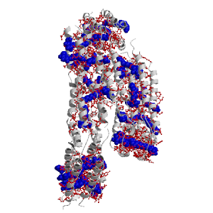
Figure showing the binding site residues. Ligands are displayed as
CPK. Figures were drawn with
Molscript (7) and rendered with
Raster3D (8). PISA coordinates
(3) are used when available
(all entries except NMR). Ligands do not appear on the picture when
PISA fails to apply symmetry operations to ligand coordinates. | | (click anywhere in this window to remove it) |
|
 |
|
List of binding site residues detected in this holo-structure. Column 1 gives the position, coloured on a yellow-to-red scale depending on the number of holo-structures where the residue is in contact with a ligand.
Column 2 gives the identifier of the chain to which the residue belongs.
Column 3 gives the 3-letter amino acid code, coloured according to physico-chemical type. The binding residues in this holo structure are listed for each ligand independently, following the ligand's unique ID. Note that since PISA files are used whenever available, chain identifiers may differ from those in original PDB files. | | (click anywhere in this window to remove it) |
|
| BOG F1625 |
|---|
| 3 | H |
LYS |
| 67 | E |
PHE |
| 70 | F |
LEU |
| 71 | E |
GLY |
| 72 | M |
THR |
| 73 | E |
GLN |
| 74 | M |
PRO |
| 75 | M |
ALA |
| 76 | M |
THR |
| BOG H1626 |
|---|
| 4 | H |
GLU |
| 7 | P |
VAL |
| 8 | P |
GLU |
| 10 | H |
LEU |
| 11 | P |
VAL |
| 12 | G |
SER |
| 14 | G |
TYR |
| 15 | G |
PHE |
| 76 | F |
THR |
| BOG J1603 |
|---|
| 4 | J |
GLU |
| 6 | J |
CYS |
| 7 | I |
VAL |
| 8 | I |
GLU |
| | 10 | B |
LEU |
| 11 | A |
VAL |
| 12 | A |
SER |
| 14 | A |
TYR |
| 15 | F |
PHE |
| BOG N1617 |
|---|
| 5 | F |
PRO |
| 6 | F |
CYS |
| 7 | N |
VAL |
| 8 | M |
GLU |
| 10 | N |
LEU |
| 11 | N |
VAL |
| 12 | N |
SER |
| 15 | N |
PHE |
| 73 | D |
GLN |
| 75 | C |
ALA |
| 76 | C |
THR |
| BOG I1601 |
|---|
| 6 | I |
CYS |
| 7 | I |
VAL |
| 8 | I |
GLU |
| 9 | I |
SER |
| 10 | J |
LEU |
| 11 | J |
VAL |
| 12 | I |
SER |
| | 13 | J |
GLN |
| 14 | J |
TYR |
| 15 | A |
PHE |
| 16 | I |
GLN |
| BOG G1627 |
|---|
| 7 | Q |
VAL |
| 11 | Q |
VAL |
| 14 | Q |
TYR |
| 18 | G |
VAL |
| BOG Q1627 |
|---|
| 7 | Q |
VAL |
| 8 | P |
GLU |
| 10 | Q |
LEU |
| 11 | Q |
VAL |
| 12 | P |
SER |
| 14 | Q |
TYR |
| 15 | P |
PHE |
| BOG C1610 |
|---|
| 7 | D |
VAL |
| 8 | C |
GLU |
| 10 | L |
LEU |
| 11 | C |
VAL |
| 12 | K |
SER |
| 13 | L |
GLN |
| 14 | L |
TYR |
| | 15 | K |
PHE |
| 16 | K |
GLN |
| 17 | L |
THR |
| 19 | K |
THR |
| BOG H1627 |
|---|
| 8 | P |
GLU |
| 10 | Q |
LEU |
| 11 | P |
VAL |
| 12 | P |
SER |
| 15 | G |
PHE |
| BOG F1619 |
|---|
| 8 | J |
GLU |
| 14 | F |
TYR |
| 15 | E |
PHE |
| 18 | N |
VAL |
| 19 | E |
THR |
| 21 | N |
TYR |
| 22 | N |
GLY |
| 23 | E |
LYS |
| 25 | N |
LEU |
| 26 | E |
MET |
| BOG E1618 |
|---|
| 8 | E |
GLU |
| 10 | N |
LEU |
| 11 | N |
VAL |
| | 14 | N |
TYR |
| 15 | M |
PHE |
| BOG N1618 |
|---|
| 8 | E |
GLU |
| 10 | N |
LEU |
| 11 | N |
VAL |
| 13 | N |
GLN |
| 14 | N |
TYR |
| 15 | E |
PHE |
| 16 | N |
GLN |
| 17 | E |
THR |
| 18 | N |
VAL |
| 19 | N |
THR |
| BOG B1601 |
|---|
| 8 | I |
GLU |
| 12 | I |
SER |
| 14 | B |
TYR |
| 15 | A |
PHE |
| BOG M1620 |
|---|
| 9 | J |
SER |
| 23 | M |
LYS |
| 24 | N |
ASP |
| 25 | M |
LEU |
| 26 | M |
MET |
| 27 | M |
GLU |
| | 28 | M |
LYS |
| 29 | M |
VAL |
| 30 | M |
LYS |
| BOG P1627 |
|---|
| 11 | P |
VAL |
| 12 | P |
SER |
| 14 | H |
TYR |
| 15 | P |
PHE |
| BOG B1602 |
|---|
| 12 | I |
SER |
| 14 | J |
TYR |
| 15 | A |
PHE |
| 17 | B |
THR |
| 18 | B |
VAL |
| 19 | A |
THR |
| 21 | B |
TYR |
| 22 | B |
GLY |
| 23 | B |
LYS |
| 26 | B |
MET |
| BOG G1628 |
|---|
| 14 | Q |
TYR |
| 15 | G |
PHE |
| 17 | H |
THR |
| 18 | G |
VAL |
| 19 | G |
THR |
| | 21 | H |
TYR |
| BOG P1631 |
|---|
| 15 | L |
PHE |
| 47 | P |
GLU |
| 48 | H |
GLN |
| 49 | G |
LEU |
| 51 | P |
PRO |
| 52 | G |
LEU |
| 53 | H |
ILE |
| 54 | P |
LYS |
| 55 | P |
LYS |
| 58 | P |
THR |
| BOG H1629 |
|---|
| 17 | Q |
THR |
| 18 | Q |
VAL |
| 19 | P |
THR |
| 21 | Q |
TYR |
| 22 | P |
GLY |
| 25 | H |
LEU |
| 26 | G |
MET |
| 29 | H |
VAL |
| BOG D1612 |
|---|
| 18 | D |
VAL |
| 21 | K |
TYR |
| 22 | D |
GLY |
| | 25 | L |
LEU |
| 26 | K |
MET |
| 28 | L |
LYS |
| 29 | C |
VAL |
| 32 | L |
PRO |
| 33 | C |
GLU |
| BOG I1605 |
|---|
| 19 | I |
THR |
| 21 | J |
TYR |
| 22 | I |
GLY |
| 24 | I |
ASP |
| 25 | B |
LEU |
| 28 | B |
LYS |
| 29 | B |
VAL |
| 32 | B |
PRO |
| 33 | B |
GLU |
| BOG A1604 |
|---|
| 21 | J |
TYR |
| 25 | B |
LEU |
| 26 | A |
MET |
| 28 | B |
LYS |
| 29 | A |
VAL |
| 30 | A |
LYS |
| 32 | A |
PRO |
| 33 | A |
GLU |
| | 36 | A |
ALA |
| 37 | A |
GLU |
| BOG B1605 |
|---|
| 25 | I |
LEU |
| 29 | B |
VAL |
| 32 | B |
PRO |
| 33 | B |
GLU |
| BOG I1606 |
|---|
| 37 | J |
GLU |
| 38 | J |
ALA |
| 40 | I |
SER |
| 41 | I |
TYR |
| 42 | I |
PHE |
| 43 | I |
GLU |
| 44 | I |
LYS |
| 45 | B |
SER |
| 48 | I |
GLN |
| BOG Q1630 |
|---|
| 37 | H |
GLU |
| 38 | Q |
ALA |
| 40 | H |
SER |
| 41 | H |
TYR |
| 42 | P |
PHE |
| BOG H1630 |
|---|
| 40 | H |
SER |
| | 41 | Q |
TYR |
| 42 | G |
PHE |
| 44 | H |
LYS |
| 45 | G |
SER |
| 48 | G |
GLN |
| BOG B1606 |
|---|
| 41 | B |
TYR |
| 42 | B |
PHE |
| 44 | I |
LYS |
| 45 | B |
SER |
| BOG C1614 |
|---|
| 41 | L |
TYR |
| 44 | D |
LYS |
| 45 | L |
SER |
| 48 | C |
GLN |
| 49 | C |
LEU |
| BOG L1614 |
|---|
| 44 | C |
LYS |
| 47 | C |
GLU |
| 48 | C |
GLN |
| 49 | L |
LEU |
| 50 | L |
THR |
| 52 | L |
LEU |
| 53 | L |
ILE |
| 54 | L |
LYS |
| | BOG J1607 |
|---|
| 47 | A |
GLU |
| 48 | B |
GLN |
| 49 | A |
LEU |
| 51 | A |
PRO |
| 52 | I |
LEU |
| 53 | J |
ILE |
| 55 | A |
LYS |
| BOG B1607 |
|---|
| 48 | I |
GLN |
| 49 | I |
LEU |
| 52 | A |
LEU |
| BOG M1621 |
|---|
| 51 | N |
PRO |
| 52 | M |
LEU |
| 53 | M |
ILE |
| 54 | N |
LYS |
| 55 | N |
LYS |
| 56 | M |
ALA |
| 57 | M |
GLY |
| 58 | N |
THR |
| 61 | M |
VAL |
| BOG A1608 |
|---|
| 58 | A |
THR |
| 60 | J |
LEU |
| | 62 | A |
ASN |
| 63 | A |
PHE |
| 66 | A |
TYR |
| BOG L1615 |
|---|
| 58 | C |
THR |
| 59 | K |
GLU |
| 60 | C |
LEU |
| 62 | C |
ASN |
| 63 | K |
PHE |
| 64 | L |
LEU |
| 66 | C |
TYR |
| 67 | L |
PHE |
| BOG B1608 |
|---|
| 59 | A |
GLU |
| 60 | A |
LEU |
| 63 | A |
PHE |
| 64 | J |
LEU |
| 66 | A |
TYR |
| 67 | A |
PHE |
| BOG G1633 |
|---|
| 59 | Q |
GLU |
| 60 | Q |
LEU |
| 62 | Q |
ASN |
| 63 | H |
PHE |
| 64 | Q |
LEU |
| | 65 | G |
SER |
| 66 | G |
TYR |
| 67 | H |
PHE |
| 69 | G |
GLU |
| BOG P1632 |
|---|
| 59 | G |
GLU |
| 60 | G |
LEU |
| 62 | P |
ASN |
| 63 | G |
PHE |
| 64 | H |
LEU |
| 65 | P |
SER |
| 66 | P |
TYR |
| 67 | P |
PHE |
| 69 | P |
GLU |
| BOG N1623 |
|---|
| 60 | N |
LEU |
| 62 | E |
ASN |
| 63 | N |
PHE |
| 64 | N |
LEU |
| 65 | N |
SER |
| 66 | E |
TYR |
| 67 | N |
PHE |
| 68 | N |
VAL |
| BOG I1609 |
|---|
| 60 | B |
LEU |
| | 62 | I |
ASN |
| 63 | J |
PHE |
| 64 | B |
LEU |
| 65 | I |
SER |
| 66 | J |
TYR |
| 67 | I |
PHE |
| 69 | I |
GLU |
| BOG M1624 |
|---|
| 62 | E |
ASN |
| 63 | E |
PHE |
| 66 | E |
TYR |
| 67 | N |
PHE |
| 68 | M |
VAL |
| 70 | E |
LEU |
| 71 | M |
GLY |
| BOG B1609 |
|---|
| 63 | J |
PHE |
| 66 | J |
TYR |
| 67 | A |
PHE |
| 70 | A |
LEU |
| BOG K1616 |
|---|
| 63 | K |
PHE |
| 64 | D |
LEU |
| 66 | K |
TYR |
| 67 | C |
PHE |
| | 70 | D |
LEU |
| 71 | K |
GLY |
| BOG G1634 |
|---|
| 64 | Q |
LEU |
| 66 | H |
TYR |
| 67 | Q |
PHE |
| 68 | Q |
VAL |
| 69 | G |
GLU |
| 70 | P |
LEU |
| 71 | Q |
GLY |
| 72 | Q |
THR |
| 73 | G |
GLN |
| BOG E1625 |
|---|
| 67 | E |
PHE |
| 70 | F |
LEU |
| 71 | E |
GLY |
|
|
|
| PDB |
The Protein Data Bank |
| CSA |
Catalytic Site Atlas |
| PDBSum |
Overview of the macromolecular structure |
| CATH |
Protein Structure Classification |
| Scop |
Structural Classification of Proteins |
| Pfam |
Protein Families and Domains |
| UniProt |
Universal Protein Resource |
LIGPLOT (only on holo-pages) is hosted at the EBI. The LigPlot Jmol links point directly to the Jmol visualisation interface provided on the PDBSum page. Note that due to different software used, the atomic contacts of LigPlot and LigASite do not necessarily correspond. | | (click anywhere in this window to remove it) |
|
Links to external databases: LigPlot (hosted at the EBI):
|
|
Several files are provided for download: | • The XML file defining the residue-ligand contacts; this file contains data on the apo and all holo-structures. |
| • The XML Schema file defining the semantics of the XML file |
| • 3D coordinates of the structure used in constructing LigASite (PISA structure file whenever available, PDB file otherwise. |
| • 3D coordinates of the combined binding residues in the apo structure |
| • 3D coordinates of the binding residues of the holo structure (only on the holo page) |
Coordinate files are in PDB format. | | (click anywhere in this window to remove it) |
|
|
|
|
|
List of related structure, containing both the apo-structure
and other holo-structures.
Column 1 gives the PDB ID and column 2 the unique ID
of the ligands (holo-structures only).
Clicking the blue 'Hide table of related structures' button
removes the entire table. | | (click anywhere in this window to remove it) |
|
|
| pdb ID |
|---|
| 2ou1 |
|
Details |
|
| pdb ID |
Ligand Unique ID |
|---|
| none |
|
|
|
Ligands present in this holo structure.
Column 1 shows the ligand HET code
Column 2 shows the name, chemical formula and (non-stereo) SMILES string.
Data in column 2 appears as 'not_found' when it is not present in the file
'pdb2smiles.xml' from www.rcsb.org. | | (click anywhere in this window to remove it) |
|
| BOG |
NAME: |
B-OCTYLGLUCOSIDE |
|
FORMULA: |
C14 H28 O6 |
|
SMILES: |
CCCCCCCCOC1OC(CO)C(O)C(O)C1O |
|
|
|
|