 |
LigASite database of binding sites |
|
PDB ID and HEADER, TITLE and
COMPND records of the PDB file. | | (click anywhere in this window to remove it) |
|
| 3kcl |
|
|
| ROOM TEMPERATURE NEUTRON STRUCTURE OF D-XYLOSE ISOMERASE IN WITH TWO CD2+ CATIONS AND D12-D-ALPHA-GLUCOSE IN THE RING F (REFINED JOINTLY WITH X-RAY STRUCTURE 3KBM) |
TITLE |
|
|
|
|
|
|
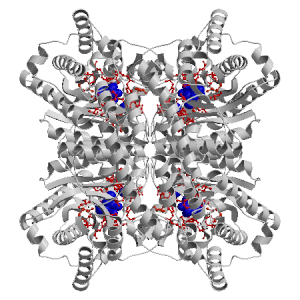
Figure showing the binding site residues. Ligands are displayed as
CPK. Figures were drawn with
Molscript (7) and rendered with
Raster3D (8). PISA coordinates
(3) are used when available
(all entries except NMR). Ligands do not appear on the picture when
PISA fails to apply symmetry operations to ligand coordinates. | | (click anywhere in this window to remove it) |
|
 |
|
List of binding site residues detected in this holo-structure. Column 1 gives the position, coloured on a yellow-to-red scale depending on the number of holo-structures where the residue is in contact with a ligand.
Column 2 gives the identifier of the chain to which the residue belongs.
Column 3 gives the 3-letter amino acid code, coloured according to physico-chemical type. The binding residues in this holo structure are listed for each ligand independently, following the ligand's unique ID. Note that since PISA files are used whenever available, chain identifiers may differ from those in original PDB files. | | (click anywhere in this window to remove it) |
|
| GLC H 401 |
|---|
| 16 | E |
TRP |
| 26 | I |
PHE |
| 54 | E |
HIS |
| 88 | E |
MET |
| 90 | E |
THR |
| 91 | E |
THR |
| 94 | E |
PHE |
| 135 | E |
VAL |
| 137 | E |
TRP |
| 181 | E |
GLU |
| 183 | E |
LYS |
| 217 | E |
GLU |
| 220 | E |
HIS |
| 245 | E |
ASP |
| | 255 | E |
ASP |
| 257 | E |
ASP |
| 287 | E |
ASP |
| 289 | E |
LYS |
| GLC P 401 |
|---|
| 16 | M |
TRP |
| 26 | A |
PHE |
| 54 | M |
HIS |
| 88 | M |
MET |
| 90 | M |
THR |
| 91 | M |
THR |
| 94 | M |
PHE |
| 135 | M |
VAL |
| 137 | M |
TRP |
| 181 | M |
GLU |
| | 183 | M |
LYS |
| 217 | M |
GLU |
| 220 | M |
HIS |
| 245 | M |
ASP |
| 255 | M |
ASP |
| 257 | M |
ASP |
| 287 | M |
ASP |
| 289 | M |
LYS |
| GLC D 401 |
|---|
| 16 | A |
TRP |
| 26 | M |
PHE |
| 54 | A |
HIS |
| 88 | A |
MET |
| 90 | A |
THR |
| 91 | A |
THR |
| | 94 | A |
PHE |
| 135 | A |
VAL |
| 137 | A |
TRP |
| 181 | A |
GLU |
| 183 | A |
LYS |
| 217 | A |
GLU |
| 220 | A |
HIS |
| 245 | A |
ASP |
| 255 | A |
ASP |
| 257 | A |
ASP |
| 287 | A |
ASP |
| 289 | A |
LYS |
| GLC L 401 |
|---|
| 16 | I |
TRP |
| 26 | E |
PHE |
| | 54 | I |
HIS |
| 88 | I |
MET |
| 90 | I |
THR |
| 91 | I |
THR |
| 94 | I |
PHE |
| 135 | I |
VAL |
| 137 | I |
TRP |
| 181 | I |
GLU |
| 183 | I |
LYS |
| 217 | I |
GLU |
| 220 | I |
HIS |
| 245 | I |
ASP |
| 255 | I |
ASP |
| 257 | I |
ASP |
| 287 | I |
ASP |
| | 289 | I |
LYS |
| CD G 392 |
|---|
| 181 | E |
GLU |
| 217 | E |
GLU |
| 245 | E |
ASP |
| 287 | E |
ASP |
| CD O 392 |
|---|
| 181 | M |
GLU |
| 217 | M |
GLU |
| 245 | M |
ASP |
| 287 | M |
ASP |
| CD C 392 |
|---|
| 181 | A |
GLU |
| 217 | A |
GLU |
| 245 | A |
ASP |
| | 287 | A |
ASP |
| CD K 392 |
|---|
| 181 | I |
GLU |
| 217 | I |
GLU |
| 245 | I |
ASP |
| 287 | I |
ASP |
| CD F 391 |
|---|
| 217 | E |
GLU |
| 220 | E |
HIS |
| 255 | E |
ASP |
| 257 | E |
ASP |
| 287 | E |
ASP |
| CD N 391 |
|---|
| 217 | M |
GLU |
| 220 | M |
HIS |
| | 255 | M |
ASP |
| 257 | M |
ASP |
| 287 | M |
ASP |
| CD B 391 |
|---|
| 217 | A |
GLU |
| 220 | A |
HIS |
| 255 | A |
ASP |
| 257 | A |
ASP |
| 287 | A |
ASP |
| CD J 391 |
|---|
| 217 | I |
GLU |
| 220 | I |
HIS |
| 255 | I |
ASP |
| 257 | I |
ASP |
| 287 | I |
ASP |
|
|
|
| PDB |
The Protein Data Bank |
| CSA |
Catalytic Site Atlas |
| PDBSum |
Overview of the macromolecular structure |
| CATH |
Protein Structure Classification |
| Scop |
Structural Classification of Proteins |
| Pfam |
Protein Families and Domains |
| UniProt |
Universal Protein Resource |
LIGPLOT (only on holo-pages) is hosted at the EBI. The LigPlot Jmol links point directly to the Jmol visualisation interface provided on the PDBSum page. Note that due to different software used, the atomic contacts of LigPlot and LigASite do not necessarily correspond. | | (click anywhere in this window to remove it) |
|
Links to external databases: LigPlot (hosted at the EBI):
|
|
Several files are provided for download: | • The XML file defining the residue-ligand contacts; this file contains data on the apo and all holo-structures. |
| • The XML Schema file defining the semantics of the XML file |
| • 3D coordinates of the structure used in constructing LigASite (PISA structure file whenever available, PDB file otherwise. |
| • 3D coordinates of the combined binding residues in the apo structure |
| • 3D coordinates of the binding residues of the holo structure (only on the holo page) |
Coordinate files are in PDB format. | | (click anywhere in this window to remove it) |
|
|
|
|
|
List of related structure, containing both the apo-structure
and other holo-structures.
Column 1 gives the PDB ID and column 2 the unique ID
of the ligands (holo-structures only).
Clicking the blue 'Hide table of related structures' button
removes the entire table. | | (click anywhere in this window to remove it) |
|
|
| pdb ID |
|---|
| 2gub |
|
Details |
|
| pdb ID |
Ligand Unique ID |
|---|
| 3kbn |
GLOO_401 _NIM_391 _NIN_393 _NIL_392 |
Details |
|
GLOT_401 _NIQ_392 _NIS_393 _NIR_391 |
|
GLOE_401 _NIB_392 _NIC_391 _NID_393 |
|
GLOJ_401 _NIG_392 _NIH_391 _NII_393 |
| 3kco |
GLOS_401 _NIT_393 _NIQ_392 _NIR_391 |
Details |
|
GLON_401 _NIO_393 _NIM_391 _NIL_392 |
|
GLOI_401 _NIG_392 _NIJ_393 _NIH_391 |
|
GLOD_401 _NIE_393 _NIB_392 _NIC_391 |
| 3kbm |
GLCH_401 _CDG_392 |
Details |
|
GLCD_401 _CDC_392 |
|
GLCP_401 _CDO_392 |
|
GLCL_401 _CDK_392 |
| 3cwh |
XULG_401 _MGI_392 _OHJ_501 _MGH_391 |
Details |
|
XULL_401 _MGN_392 _MGM_391 _OHO_501 |
|
XULQ_401 _OHT_501 _MGS_392 _MGR_391 |
|
XULB_401 _MGC_391 _MGD_392 _OHE_501 |
|
|
|
Ligands present in this holo structure.
Column 1 shows the ligand HET code
Column 2 shows the name, chemical formula and (non-stereo) SMILES string.
Data in column 2 appears as 'not_found' when it is not present in the file
'pdb2smiles.xml' from www.rcsb.org. | | (click anywhere in this window to remove it) |
|
| GLC |
NAME: |
GLUCOSE |
|
FORMULA: |
C6 H12 O6 |
|
SMILES: |
OCC1OC(O)C(O)C(O)C1O |
| CD |
NAME: |
CADMIUM ION |
|
FORMULA: |
CD1 |
|
SMILES: |
[Cd++] |
|
|
|
|
v9.0
September 2010 |
Interdisciplinary Research Institute, Computational Biology, Villeneuve d'Ascq, France
University College London, Biomolecular Structure and Modelling Unit, London, UK
Hospital for Sick Children and University of Toronto, Structural Biology and Biochemistry Program, Toronto, Canada |
| Script execution time: 0.5949 seconds |