 |
LigASite database of binding sites |
|
PDB ID and HEADER, TITLE and
COMPND records of the PDB file. | | (click anywhere in this window to remove it) |
|
| 1cim |
|
|
| THE POSITIONS OF HIS-64 AND A BOUND WATER IN HUMAN CARBONIC ANHYDRASE II UPON BINDING THREE STRUCTURALLY RELATED INHIBITORS |
TITLE |
|
|
| CARBONIC ANHYDRASE II |
COMPND |
|
|
|
|
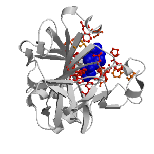
Figure showing the binding site residues. Ligands are displayed as
CPK. Figures were drawn with
Molscript (7) and rendered with
Raster3D (8). PISA coordinates
(3) are used when available
(all entries except NMR). Ligands do not appear on the picture when
PISA fails to apply symmetry operations to ligand coordinates. | | (click anywhere in this window to remove it) |
|
 |
|
List of binding site residues detected in this holo-structure. Column 1 gives the position, coloured on a yellow-to-red scale depending on the number of holo-structures where the residue is in contact with a ligand.
Column 2 gives the identifier of the chain to which the residue belongs.
Column 3 gives the 3-letter amino acid code, coloured according to physico-chemical type. The binding residues in this holo structure are listed for each ligand independently, following the ligand's unique ID. Note that since PISA files are used whenever available, chain identifiers may differ from those in original PDB files. | | (click anywhere in this window to remove it) |
|
| PTS 262 |
|---|
| 5 | |
TRP |
| 62 | |
ASN |
| 64 | |
HIS |
| 91 | |
ILE |
| 92 | |
GLN |
| 94 | |
HIS |
| 96 | |
HIS |
| 119 | |
HIS |
| 121 | |
VAL |
| 131 | |
PHE |
| 135 | |
VAL |
| 141 | |
LEU |
| 143 | |
VAL |
| 199 | |
THR |
| 201 | |
PRO |
| 202 | |
PRO |
| 209 | |
TRP |
| ZN 261 |
|---|
| 94 | |
HIS |
| 96 | |
HIS |
| 119 | |
HIS |
|
|
|
| PDB |
The Protein Data Bank |
| CSA |
Catalytic Site Atlas |
| PDBSum |
Overview of the macromolecular structure |
| CATH |
Protein Structure Classification |
| Scop |
Structural Classification of Proteins |
| Pfam |
Protein Families and Domains |
| UniProt |
Universal Protein Resource |
LIGPLOT (only on holo-pages) is hosted at the EBI. The LigPlot Jmol links point directly to the Jmol visualisation interface provided on the PDBSum page. Note that due to different software used, the atomic contacts of LigPlot and LigASite do not necessarily correspond. | | (click anywhere in this window to remove it) |
|
Links to external databases: LigPlot (hosted at the EBI):
|
|
Several files are provided for download: | • The XML file defining the residue-ligand contacts; this file contains data on the apo and all holo-structures. |
| • The XML Schema file defining the semantics of the XML file |
| • 3D coordinates of the structure used in constructing LigASite (PISA structure file whenever available, PDB file otherwise. |
| • 3D coordinates of the combined binding residues in the apo structure |
| • 3D coordinates of the binding residues of the holo structure (only on the holo page) |
Coordinate files are in PDB format. | | (click anywhere in this window to remove it) |
|
|
|
|
|
List of related structure, containing both the apo-structure
and other holo-structures.
Column 1 gives the PDB ID and column 2 the unique ID
of the ligands (holo-structures only).
Clicking the blue 'Hide table of related structures' button
removes the entire table. | | (click anywhere in this window to remove it) |
|
|
| pdb ID |
|---|
| 2cbe |
|
Details |
|
| pdb ID |
Ligand Unique ID |
|---|
| 1okl |
MNS__862 _ZN__262 |
Details |
| 1if9 |
SBBB_555 _ZNA_262 |
Details |
| 1oq5 |
CELB_701 _ZNA_600 |
Details |
| 1if8 |
SBSB_555 _ZNA_262 |
Details |
| 2hl4 |
BO1B_264 _ZNA_262 GOLB_265 |
Details |
| 1zge |
GOLB_900 SDAB_300 _ZNB1000 |
Details |
| 1xq0 |
4TRB_270 _ZNA_262 |
Details |
| 1cin |
MTS__262 _ZN__261 |
Details |
| 1cil |
ETS__262 _ZN__261 |
Details |
| 1i8z |
INLB_555 _ZNA_262 |
Details |
| 1okn |
STB__555 _ZN__262 |
Details |
| 2aw1 |
COXB_264 GOLB_266 _ZNB_262 |
Details |
| 1ze8 |
GOLB_700 _ZNA_263 PIUA___1 |
Details |
| 1z9y |
FUNB_500 _ZNB_300 |
Details |
| 2qoa |
GOLA_802 _ZNA_262 MAJA_800 |
Details |
| 2gd8 |
PO1B_264 PO1B_265 _ZNB_262 |
Details |
| 1lug |
GOLB1003 _ZNA1001 SUAA1002 _HGA1005 |
Details |
| 2hoc |
1CNB_266 MBOA_267 |
Details |
|
1CNB_265 _ZNB_263 GOLB_702 |
| 1zfq |
GOLB_900 _ZNB_600 ZECB_300 |
Details |
| 1okm |
SAB__555 _ZN__262 |
Details |
| 1ttm |
667B_264 _ZNB_262 |
Details |
| 1if7 |
SBRB_555 _ZNA_262 |
Details |
| 1i90 |
INMB_555 _ZNA_262 |
Details |
| 2qp6 |
MB1A_800 _ZNA_262 |
Details |
| 2qo8 |
3CCA_800 _ZNA_262 GOLA_802 |
Details |
| 2hd6 |
BOSB_266 GOLB_264 _ZNB_262 |
Details |
| 1i91 |
INQB_555 _ZNA_262 |
Details |
| 1zgf |
TRUB_300 _ZNB_400 |
Details |
| 2f14 |
FL1B1270 GOLB1266 _ZNB1262 GOLB1267 |
Details |
| 1zfk |
GOLB1900 NR2B1400 _ZNB1300 |
Details |
|
NR2B1500 |
|
|
|
Ligands present in this holo structure.
Column 1 shows the ligand HET code
Column 2 shows the name, chemical formula and (non-stereo) SMILES string.
Data in column 2 appears as 'not_found' when it is not present in the file
'pdb2smiles.xml' from www.rcsb.org. | | (click anywhere in this window to remove it) |
|
| PTS |
NAME: |
(4S-TRANS)-4-(AMINO)-5,6-DIHYDRO-6-METHYL-4H-THIENO (2,3-B)THIOPYRAN-2-SULFONAMIDE-7,7-DIOXIDE |
|
FORMULA: |
C8 H12 N2 O4 S3 |
|
SMILES: |
CC1CC(N)c2cc(sc2S1(=O)=O)S(N)(=O)=O |
| ZN |
NAME: |
ZINC ION |
|
FORMULA: |
ZN1 |
|
SMILES: |
[Zn++] |
|
|
|
|
v6.0
October 2008 |
Interdisciplinary Research Institute, Computational Biology, Villeneuve d'Ascq, France
University College London, Biomolecular Structure and Modelling Unit, London, UK
Hospital for Sick Children and University of Toronto, Structural Biology and Biochemistry Program, Toronto, Canada |
| Script execution time: 1.1109 seconds |