 |
LigASite database of binding sites |
|
PDB ID and HEADER, TITLE and
COMPND records of the PDB file. | | (click anywhere in this window to remove it) |
|
| 1tt6 |
|
|
| THE ORTHORHOMBIC CRYSTAL STRUCTURE OF TRANSTHYRETIN IN COMPLEX WITH DIETHYLSTILBESTROL |
TITLE |
|
|
|
|
|
|
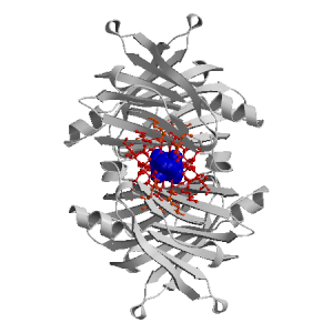
Figure showing the binding site residues. Ligands are displayed as
CPK. Figures were drawn with
Molscript (7) and rendered with
Raster3D (8). PISA coordinates
(3) are used when available
(all entries except NMR). Ligands do not appear on the picture when
PISA fails to apply symmetry operations to ligand coordinates. | | (click anywhere in this window to remove it) |
|
 |
|
List of binding site residues detected in this holo-structure. Column 1 gives the position, coloured on a yellow-to-red scale depending on the number of holo-structures where the residue is in contact with a ligand.
Column 2 gives the identifier of the chain to which the residue belongs.
Column 3 gives the 3-letter amino acid code, coloured according to physico-chemical type. The binding residues in this holo structure are listed for each ligand independently, following the ligand's unique ID. Note that since PISA files are used whenever available, chain identifiers may differ from those in original PDB files. | | (click anywhere in this window to remove it) |
|
| DES E 2 |
|---|
| 13 | C |
MET |
| 15 | A |
LYS |
| 17 | A |
LEU |
| 106 | C |
THR |
| 108 | A |
ALA |
| 109 | A |
ALA |
| 110 | A |
LEU |
| 117 | C |
SER |
| 118 | C |
THR |
| 119 | C |
THR |
| 121 | C |
VAL |
| DES F 2 |
|---|
| 13 | A |
MET |
| 15 | A |
LYS |
| | 17 | C |
LEU |
| 54 | A |
GLU |
| 106 | A |
THR |
| 108 | A |
ALA |
| 109 | C |
ALA |
| 110 | C |
LEU |
| 117 | A |
SER |
| 118 | A |
THR |
| 119 | A |
THR |
| 121 | A |
VAL |
| DES E 1 |
|---|
| 15 | B |
LYS |
| 17 | B |
LEU |
| 106 | B |
THR |
| 108 | B |
ALA |
| | 109 | B |
ALA |
| 110 | B |
LEU |
| 117 | D |
SER |
| 118 | D |
THR |
| 119 | D |
THR |
| 121 | B |
VAL |
| DES F 1 |
|---|
| 15 | D |
LYS |
| 17 | D |
LEU |
| 106 | D |
THR |
| 108 | D |
ALA |
| 109 | D |
ALA |
| 110 | D |
LEU |
| 117 | B |
SER |
| 118 | B |
THR |
| |
|
|
| PDB |
The Protein Data Bank |
| CSA |
Catalytic Site Atlas |
| PDBSum |
Overview of the macromolecular structure |
| CATH |
Protein Structure Classification |
| Scop |
Structural Classification of Proteins |
| Pfam |
Protein Families and Domains |
| UniProt |
Universal Protein Resource |
LIGPLOT (only on holo-pages) is hosted at the EBI. The LigPlot Jmol links point directly to the Jmol visualisation interface provided on the PDBSum page. Note that due to different software used, the atomic contacts of LigPlot and LigASite do not necessarily correspond. | | (click anywhere in this window to remove it) |
|
Links to external databases: LigPlot (hosted at the EBI):
|
|
Several files are provided for download: | • The XML file defining the residue-ligand contacts; this file contains data on the apo and all holo-structures. |
| • The XML Schema file defining the semantics of the XML file |
| • 3D coordinates of the structure used in constructing LigASite (PISA structure file whenever available, PDB file otherwise. |
| • 3D coordinates of the combined binding residues in the apo structure |
| • 3D coordinates of the binding residues of the holo structure (only on the holo page) |
Coordinate files are in PDB format. | | (click anywhere in this window to remove it) |
|
|
|
|
|
List of related structure, containing both the apo-structure
and other holo-structures.
Column 1 gives the PDB ID and column 2 the unique ID
of the ligands (holo-structures only).
Clicking the blue 'Hide table of related structures' button
removes the entire table. | | (click anywhere in this window to remove it) |
|
|
| pdb ID |
|---|
| 1f41 |
|
Details |
|
| pdb ID |
Ligand Unique ID |
|---|
| 1ict |
T44A_128 T44C_128 |
Details |
|
T44B_129 T44D_129 |
| 2f7i |
26CE_326 26CF_326 |
Details |
|
26CE_325 26CF_325 |
| 1z7j |
T4AE_129 T4AF_129 |
Details |
|
T4AE_128 T4AF_128 |
| 1y1d |
FHIE3000 FHIF3000 |
Details |
|
FHIE2000 FHIF2000 |
| 1tz8 |
DESC___1 DESD___1 |
Details |
|
DESA___1 DESB___1 |
| 1eta |
T44C_128 T44D_128 |
Details |
| 2qgd |
MR5B_201 MR5D_201 |
Details |
|
MR5A_200 MR5C_200 |
| 2b77 |
3CAE_240 3CAF_240 |
Details |
|
3CAE_239 3CAF_239 |
| 3b56 |
DIUB3000 DIUD3000 |
Details |
|
DIUA2000 DIUC2000 |
| 1u21 |
P2CE_211 P2CF_211 |
Details |
|
P2CE_221 P2CF_221 |
| 2fbr |
44CE_173 44CF_173 |
Details |
| 3d2t |
1FLB_500 1FLD_500 |
Details |
|
1FLA_502 1FLC_502 |
| 2b15 |
DNFE___2 DNFF___2 |
Details |
|
DNFE___1 DNFF___1 |
| 2roy |
P28B_128 P28D_128 |
Details |
|
P28A_128 P28C_128 |
| 1bm7 |
FLFE_502 FLFF_502 |
Details |
|
FLFE_501 FLFF_501 |
| 2gab |
NE2E_502 NE2F_502 |
Details |
|
NE2E_501 NE2F_501 |
| 2f8i |
205E1002 205F1002 |
Details |
|
205E1001 205F1001 |
| 2qge |
MR6A_201 MR6C_201 |
Details |
|
MR6B_200 MR6D_200 |
| 2b9a |
FBCE_301 FBCF_301 |
Details |
|
FBCE_302 FBCF_302 |
| 2flm |
6CAE_201 6CAF_201 |
Details |
| 2g9k |
NE1E_500 NE1F_500 |
Details |
|
NE1E_501 NE1F_501 |
| 2qgc |
MR4B_128 MR4D_128 |
Details |
|
MR4A_128 MR4C_128 |
| 2g5u |
NEWE_240 NEWF_240 |
Details |
|
NEWE_239 NEWF_239 |
| 2rox |
T44A_128 T44C_128 |
Details |
|
T44B_128 T44D_128 |
|
|
|
Ligands present in this holo structure.
Column 1 shows the ligand HET code
Column 2 shows the name, chemical formula and (non-stereo) SMILES string.
Data in column 2 appears as 'not_found' when it is not present in the file
'pdb2smiles.xml' from www.rcsb.org. | | (click anywhere in this window to remove it) |
|
| DES |
NAME: |
DIETHYLSTILBESTROL |
|
FORMULA: |
C18 H20 O2 |
|
SMILES: |
CCC(c1ccc(O)cc1)=C(CC)c2ccc(O)cc2 |
|
|
|
|
v6.0
October 2008 |
Interdisciplinary Research Institute, Computational Biology, Villeneuve d'Ascq, France
University College London, Biomolecular Structure and Modelling Unit, London, UK
Hospital for Sick Children and University of Toronto, Structural Biology and Biochemistry Program, Toronto, Canada |
| Script execution time: 3.4223 seconds |