 |
LigASite database of binding sites |
|
PDB ID and HEADER, TITLE and
COMPND records of the PDB file. | | (click anywhere in this window to remove it) |
|
| 1w1v |
|
|
| CRYSTAL STRUCTURE OF S. MARCESCENS CHITINASE B IN COMPLEX WITH THE CYCLIC DIPEPTIDE INHIBITOR CYCLO-(L-ARG-L-PRO) AT 1.85 A RESOLUTION |
TITLE |
|
|
|
|
|
|
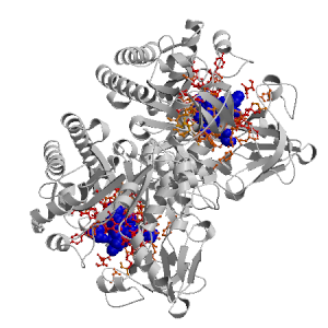
Figure showing the binding site residues. Ligands are displayed as
CPK. Figures were drawn with
Molscript (7) and rendered with
Raster3D (8). PISA coordinates
(3) are used when available
(all entries except NMR). Ligands do not appear on the picture when
PISA fails to apply symmetry operations to ligand coordinates. | | (click anywhere in this window to remove it) |
|
 |
|
List of binding site residues detected in this holo-structure. Column 1 gives the position, coloured on a yellow-to-red scale depending on the number of holo-structures where the residue is in contact with a ligand.
Column 2 gives the identifier of the chain to which the residue belongs.
Column 3 gives the 3-letter amino acid code, coloured according to physico-chemical type. The binding residues in this holo structure are listed for each ligand independently, following the ligand's unique ID. Note that since PISA files are used whenever available, chain identifiers may differ from those in original PDB files. | | (click anywhere in this window to remove it) |
|
| ALJ T1520 |
|---|
| 10 | A |
TYR |
| 51 | A |
PHE |
| 96 | A |
GLY |
| 97 | A |
TRP |
| 142 | A |
ASP |
| 144 | A |
GLU |
| 184 | A |
ALA |
| 212 | A |
MET |
| 214 | A |
TYR |
| 215 | A |
ASP |
| 292 | A |
TYR |
| 294 | A |
ARG |
| 336 | A |
ASP |
| 339 | A |
ILE |
| | 401 | A |
MET |
| 403 | A |
TRP |
| 407 | A |
GLN |
| ALJ K1513 |
|---|
| 10 | V |
TYR |
| 51 | V |
PHE |
| 96 | V |
GLY |
| 97 | V |
TRP |
| 142 | V |
ASP |
| 144 | V |
GLU |
| 184 | V |
ALA |
| 212 | V |
MET |
| 214 | V |
TYR |
| 215 | V |
ASP |
| 292 | V |
TYR |
| | 294 | V |
ARG |
| 316 | V |
ASP |
| 339 | V |
ILE |
| 401 | V |
MET |
| 403 | V |
TRP |
| 407 | V |
GLN |
| GOL F1509 |
|---|
| 12 | A |
PHE |
| 51 | A |
PHE |
| 95 | A |
GLY |
| 96 | A |
GLY |
| 97 | A |
TRP |
| 98 | A |
TYR |
| 403 | A |
TRP |
| | GOL J1517 |
|---|
| 97 | A |
TRP |
| 191 | A |
PHE |
| 215 | A |
ASP |
| 220 | A |
TRP |
| 481 | V |
TYR |
| GOL R1516 |
|---|
| 97 | A |
TRP |
| 144 | A |
GLU |
| 212 | A |
MET |
| 215 | A |
ASP |
| 220 | A |
TRP |
| 294 | A |
ARG |
| 316 | A |
ASP |
| | GOL B1512 |
|---|
| 97 | V |
TRP |
| 191 | V |
PHE |
| 220 | V |
TRP |
| 314 | V |
GLY |
| 316 | V |
ASP |
| 481 | A |
TYR |
| GOL H1510 |
|---|
| 97 | V |
TRP |
| 191 | V |
PHE |
| 215 | V |
ASP |
| 220 | V |
TRP |
| 294 | V |
ARG |
| GOL X1511 |
|---|
| 97 | V |
TRP |
| | 144 | V |
GLU |
| 145 | V |
TYR |
| 191 | V |
PHE |
| 212 | V |
MET |
| 215 | V |
ASP |
| GOL Y1508 |
|---|
| 145 | V |
TYR |
| 186 | V |
ALA |
| 187 | V |
GLY |
| 188 | V |
GLY |
| 190 | V |
PHE |
| 191 | V |
PHE |
| 212 | V |
MET |
| 214 | V |
TYR |
| 215 | V |
ASP |
| | 216 | V |
LEU |
| GOL E1500 |
|---|
| 190 | A |
PHE |
| 220 | A |
TRP |
| 221 | A |
GLU |
| 239 | A |
PHE |
| 464 | V |
GLN |
| 479 | V |
TRP |
| 480 | V |
GLY |
| 481 | V |
TYR |
| GOL K1506 |
|---|
| 190 | A |
PHE |
| 191 | A |
PHE |
| 194 | A |
ARG |
| 479 | V |
TRP |
| | 480 | V |
GLY |
| 481 | V |
TYR |
| 482 | V |
ILE |
| 483 | V |
THR |
| 484 | V |
SER |
| 489 | V |
ASP |
| 490 | V |
SER |
| 491 | V |
ALA |
| GOL D1500 |
|---|
| 190 | V |
PHE |
| 191 | V |
PHE |
| 193 | V |
SER |
| 194 | V |
ARG |
| 479 | A |
TRP |
| 480 | A |
GLY |
| | 481 | A |
TYR |
| 482 | A |
ILE |
| 483 | A |
THR |
| 489 | A |
ASP |
| GOL Z1507 |
|---|
| 190 | V |
PHE |
| 220 | V |
TRP |
| 221 | V |
GLU |
| 239 | V |
PHE |
| 464 | A |
GLN |
| 479 | A |
TRP |
| 480 | A |
GLY |
| 481 | A |
TYR |
|
|
|
| PDB |
The Protein Data Bank |
| CSA |
Catalytic Site Atlas |
| PDBSum |
Overview of the macromolecular structure |
| CATH |
Protein Structure Classification |
| Scop |
Structural Classification of Proteins |
| Pfam |
Protein Families and Domains |
| UniProt |
Universal Protein Resource |
LIGPLOT (only on holo-pages) is hosted at the EBI. The LigPlot Jmol links point directly to the Jmol visualisation interface provided on the PDBSum page. Note that due to different software used, the atomic contacts of LigPlot and LigASite do not necessarily correspond. | | (click anywhere in this window to remove it) |
|
Links to external databases: LigPlot (hosted at the EBI):
|
|
Several files are provided for download: | • The XML file defining the residue-ligand contacts; this file contains data on the apo and all holo-structures. |
| • The XML Schema file defining the semantics of the XML file |
| • 3D coordinates of the structure used in constructing LigASite (PISA structure file whenever available, PDB file otherwise. |
| • 3D coordinates of the combined binding residues in the apo structure |
| • 3D coordinates of the binding residues of the holo structure (only on the holo page) |
Coordinate files are in PDB format. | | (click anywhere in this window to remove it) |
|
|
|
|
|
List of related structure, containing both the apo-structure
and other holo-structures.
Column 1 gives the PDB ID and column 2 the unique ID
of the ligands (holo-structures only).
Clicking the blue 'Hide table of related structures' button
removes the entire table. | | (click anywhere in this window to remove it) |
|
|
| pdb ID |
|---|
| 1e15 |
|
Details |
|
| pdb ID |
Ligand Unique ID |
|---|
| 1e6r |
AMIL1502 NAAK1501 NAAJ1500 |
Details |
|
AMIF1501 NAAE1500 NAAD1499 |
| 1w1p |
GIOL1518 GOLI1510 GOLK1514 |
Details |
|
GOLH1508 GOLV1512 |
| 1ur8 |
GDLI2502 NAGH2501 |
Details |
|
GDLP2502 SO4L1510 NAGO2501 |
| 1w1y |
TYPJ1508 TYPQ1506 TYPN1507 |
Details |
|
GOLS1504 GOLU1503 |
|
TYPD1508 TYPR1509 TYPI1507 |
| 1w1t |
GOLC1506 GOLP1500 |
Details |
|
CHQB1513 CHQY1512 GOLX1507 |
|
CHQI1513 GOLF1508 CHQM1514 GOLG1509 GOLO1500 |
| 1o6i |
ARGU1520 GOLZ1510 GOLL1513 GOLJ1499 DPRU1519 GOLN1508 GOLH1515 GOLD1512 |
Details |
|
ARGA1517 GOLG1511 DPRA1516 |
|
|
|
Ligands present in this holo structure.
Column 1 shows the ligand HET code
Column 2 shows the name, chemical formula and (non-stereo) SMILES string.
Data in column 2 appears as 'not_found' when it is not present in the file
'pdb2smiles.xml' from www.rcsb.org. | | (click anywhere in this window to remove it) |
|
| GOL |
NAME: |
GLYCEROL |
|
FORMULA: |
C3 H8 O3 |
|
SMILES: |
OCC(O)CO |
| ALJ |
NAME: |
CYCLO-(L-ARGININE-L-PROLINE) INHIBITOR |
|
FORMULA: |
C11 H19 N5 O2 |
|
SMILES: |
NC(N)=NCCCC1NC(=O)C2CCCN2C1=O |
|
|
|
|
v9.7
April 2012 |
Interdisciplinary Research Institute, Computational Biology, Villeneuve d'Ascq, France
University College London, Biomolecular Structure and Modelling Unit, London, UK
Hospital for Sick Children and University of Toronto, Structural Biology and Biochemistry Program, Toronto, Canada |
| Script execution time: 1.0654 seconds |