 |
LigASite database of binding sites |
|
PDB ID and HEADER, TITLE and
COMPND records of the PDB file. | | (click anywhere in this window to remove it) |
|
| 1hon |
| LIGASE (SYNTHETASE) |
HEADER |
|
|
| STRUCTURE OF GUANINE NUCLEOTIDE (GPPCP) COMPLEX OF ADENYLOSUCCINATE SYNTHETASE FROM ESCHERICHIA COLI AT PH 6.5 AND 25 DEGREE CELSIUS |
TITLE |
|
|
| ADENYLOSUCCINATE SYNTHETASE |
COMPND |
|
|
|
|
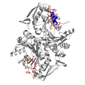
Figure showing the binding site residues. Ligands are displayed as
CPK. Figures were drawn with
Molscript (7) and rendered with
Raster3D (8). PISA coordinates
(3) are used when available
(all entries except NMR). Ligands do not appear on the picture when
PISA fails to apply symmetry operations to ligand coordinates. | | (click anywhere in this window to remove it) |
|
 |
|
List of binding site residues detected in this holo-structure. Column 1 gives the position, coloured on a yellow-to-red scale depending on the number of holo-structures where the residue is in contact with a ligand.
Column 2 gives the identifier of the chain to which the residue belongs.
Column 3 gives the 3-letter amino acid code, coloured according to physico-chemical type. The binding residues in this holo structure are listed for each ligand independently, following the ligand's unique ID. Note that since PISA files are used whenever available, chain identifiers may differ from those in original PDB files. | | (click anywhere in this window to remove it) |
|
| GNH C 432 |
|---|
| 13 | A |
ASP |
| 15 | A |
GLY |
| 16 | A |
LYS |
| 17 | A |
GLY |
| 18 | A |
LYS |
| 19 | A |
ILE |
| 40 | A |
GLY |
| 41 | A |
HIS |
| 42 | A |
THR |
| 293 | A |
GLN |
| 294 | A |
GLY |
| 300 | A |
THR |
| 330 | A |
THR |
| 331 | A |
LYS |
| | 333 | A |
ASP |
| 334 | A |
VAL |
| 383 | A |
PHE |
| 414 | A |
SER |
| 416 | A |
GLY |
| 417 | A |
PRO |
|
|
|
| PDB |
The Protein Data Bank |
| CSA |
Catalytic Site Atlas |
| PDBSum |
Overview of the macromolecular structure |
| CATH |
Protein Structure Classification |
| Scop |
Structural Classification of Proteins |
| Pfam |
Protein Families and Domains |
| UniProt |
Universal Protein Resource |
LIGPLOT (only on holo-pages) is hosted at the EBI. The LigPlot Jmol links point directly to the Jmol visualisation interface provided on the PDBSum page. Note that due to different software used, the atomic contacts of LigPlot and LigASite do not necessarily correspond. | | (click anywhere in this window to remove it) |
|
Links to external databases: LigPlot (hosted at the EBI):
|
|
Several files are provided for download: | • The XML file defining the residue-ligand contacts; this file contains data on the apo and all holo-structures. |
| • The XML Schema file defining the semantics of the XML file |
| • 3D coordinates of the structure used in constructing LigASite (PISA structure file whenever available, PDB file otherwise. |
| • 3D coordinates of the combined binding residues in the apo structure |
| • 3D coordinates of the binding residues of the holo structure (only on the holo page) |
Coordinate files are in PDB format. | | (click anywhere in this window to remove it) |
|
|
|
|
|
List of related structure, containing both the apo-structure
and other holo-structures.
Column 1 gives the PDB ID and column 2 the unique ID
of the ligands (holo-structures only).
Clicking the blue 'Hide table of related structures' button
removes the entire table. | | (click anywhere in this window to remove it) |
|
|
| pdb ID |
|---|
| 1ade |
|
Details |
|
| pdb ID |
Ligand Unique ID |
|---|
| 1hop |
GCPA_432 |
Details |
|
GCPB_433 |
| 2gcq |
DOIC_451 HADC_437 GDPC_432 _MGA_435 |
Details |
|
DOID_451 GDPD_432 _MGB_435 HADD_437 |
| 1soo |
HNPB_453 SO4B_443 _NAB_547 SO4B_442 |
Details |
|
HNPA_453 SO4A_443 SO4A_442 _NAA_547 |
| 1hoo |
GNHC_432 GNPC_432 |
Details |
| 1son |
AMPB_453 |
Details |
|
AMPA_453 |
| 1nht |
GDPB_432 _MGB_435 HADB_437 SPGB_440 |
Details |
|
GDPA_432 HADA_437 SPGA_440 _MGA_435 |
| 1cib |
GDPD_432 IMPD_440 _MGD_434 HDAD_437 NO3D_433 |
Details |
|
GDPC_432 _MGC_434 NO3C_433 IMPC_440 HDAC_437 |
| 1gim |
GDPA_432 NO3A_433 IMPA_440 HADA_438 _MGA_435 |
Details |
|
GDPB_432 IMPB_440 NO3B_433 _MGB_435 HADB_438 |
| 1gin |
GDPA_432 NO3A_433 IMPA_440 HADA_438 _MGA_435 |
Details |
|
GDPB_432 IMPB_440 NO3B_433 _MGB_435 HADB_438 |
| 1qf5 |
GDPD___1 PO4D___2 _MGD___3 RPLD___4 |
Details |
|
GDPC___1 PO4C___2 _MGC___3 RPLC___4 |
| 1ksz |
GDPA_432 HADA_437 PGSA_440 _MGA_435 |
Details |
|
GDPB_432 PGSB_440 _MGB_435 HADB_437 |
| 1qf4 |
GDPC___1 PO4C___2 RPDC___4 _MGC___3 |
Details |
|
GDPD___1 PO4D___2 _MGD___3 RPDD___4 |
| 1ch8 |
GPXD_432 IMPD_440 _MGD_434 HDAD_437 NO3D_433 |
Details |
|
GPXC_432 _MGC_434 NO3C_433 IMPC_440 HDAC_437 |
| 1juy |
GDPA_432 _PIA_434 H5PA_433 HDAA_438 _MGA_435 |
Details |
|
GDPB_432 _PIB_434 _MGB_435 HDAB_438 H5PB_433 |
|
|
|
Ligands present in this holo structure.
Column 1 shows the ligand HET code
Column 2 shows the name, chemical formula and (non-stereo) SMILES string.
Data in column 2 appears as 'not_found' when it is not present in the file
'pdb2smiles.xml' from www.rcsb.org. | | (click anywhere in this window to remove it) |
|
| GNH |
NAME: |
AMINOPHOSPHONIC ACID-GUANYLATE ESTER |
|
FORMULA: |
C10 H16 N6 O10 P2 |
|
SMILES: |
NC1=Nc2[n](cnc2C(=O)N1)C3OC(COP(O)(=O)OP(N)(O)=O)C(O)C3O |
|
|
|
|
v6.0
October 2008 |
Interdisciplinary Research Institute, Computational Biology, Villeneuve d'Ascq, France
University College London, Biomolecular Structure and Modelling Unit, London, UK
Hospital for Sick Children and University of Toronto, Structural Biology and Biochemistry Program, Toronto, Canada |
| Script execution time: 2.5128 seconds |